|
|
| |
|
 |
|
| |
 HOME > Cell Biology > Cell signaling |
|
|

|
ATP Assay Kit |
| |
|
|
|
|
|
 |
코 드 |
: 21609 |
|
 |
단 위 |
: 1 kit |
|
 |
공 급 원 |
: AAT Bioquest |
|
 |
가 격 |
: 문의 |
 |
다운로드 |
|
|
|
|
|
|
|
| |
|
|
|
| |
 |
|
 |
| |
|
|
|
| |
 |
| |
ATP Assay Kit
PhosphoWorks™ Luminometric ATP Assay Kit *Steady Glow*
ATP plays a fundamental role in cellular energenics, metabolic regulation and cellular signaling. ATP Assay Kit provides a fast, simple and homogeneous luminescence assay for the determination of cell proliferation and cytotoxicity in mammalian cells. The assay can be performed in a convenient 96-well and 384-well microtiter-plate format. The high sensitivity of this assay permits the detection of ATP in many biological systems, environmental samples and foods. This Phospho Works ATP Assay Kit has the stable luminescence signal as long as 4 hours. It has stable luminescence with no mixing or separations required, and formulated to have minimal hands-on time.
Key Features
Sensitive : Can detect as low as 10 cells/well.
Continuous : Stable luminescence, suitable for manual or automated operations with no mixing
or Separations required.
Convenient : Formulated to have minimal hands-on time.
Non-Radioactive: No special requirements for waste treatment.
Kit Components
Component A : ATP monitoring enzyme 1 vial
Component B : ATP sensor (light-sensitive) 1 vial
Component C : Reaction buffer 1 vial (10 ml)
Brief Summary
→ Prepare cells (samples) with test compounds (100 μL /96-well-plate or /25 μL96-well-plate)
→ Add equal volume of ATP assay solution
→ Incubate at room temperature for 10-20 min
→ Read luminescence

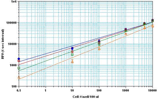
Fig 1. CHO-K1 cell number response on 96-well using NOVOstar with ABD ATP glow kit. The linear
luminescence signal for CHO-K1 cells down to 10 cells per well was detected up to 2 hr (Z’ factor = 0.6).
The integrated time was 1 sec. The half life is more than 1.5 hours

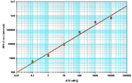
Fig 2. ATP dose response on 96-well using NOVOstar with PhosphoWorks™ ATP Assay Kit *Steady
Glow*. The linear luminescence signal for ATP concentrations from 10 uM to 0.1 nM was detected up to 5
hr (Z’ factor = 0.7) without signal decayed (above fig shows 20 min, 1, 2, 3, 4, and 5 hr signal).
The integrated time was 1 sec.
References
1. McElroy, W.D. (1947) The Energy Source for Bioluminescence in an isolated System. Proc. Natl.Acad. Sci. USA
33,342.
2. de Wet JR, Wood KV, Helinski DR, DeLuca M, (1985) Cloning of firefly luciferase cDNA and the expression of active
luciferase in Escherichia coli, Proc. Natl. Acad. Sci USA 82,7870-7873.
3. Khan, H.A. (2003) Bioluminometric assay of ATP in mouse brain: Determinant factors for enhanced test sensitivity,
J. Bioscience 28, 379-382.
4. Drew, B and C. Leeuwenburgh (2003) Method for measuring ATP production in isolated mitochondria:
ATP production in brain and liver mitochondria fo Fischer-344 rats with age and caloric restriction,
Am J. Physiol. Regul. Integr. Comp. Physiol., 285, R1260-R1268.
5. Hara, K. Y. and Mori, H. (2006) An efficient method for quantitative determination of cellular ATP synthetic activity,
J Biomol Screen 11, 310-7.
6. Sun, Y. and Chai, T. C. (2006) Augmented extracellular ATP signaling in bladder urothelial cells from patients with
interstitial cystitis Am J Physiol Cell Physiol 290, C27-34 |
| |
|
| |
 |
| |
ATP Assay
|
Product Code |
Unit |
Price |
Availability |
|
21609 |
1 x 96 well plate |
|
|
|
| |
|
|


